Dog Elephant Skin? 5 Causes Not Just Yeast + Vet Decision
Elephant skin on dogs (lichenification) — 5 causes (yeast / atopic / endocrine / hyperkeratosis / mange) identification + vet decision framework. Not just yeast.
Published 2026-06-19
Dog Elephant Skin — Need a Faster Differential?
Upload a clear photo of your dog's thickened darkened skin for an instant AI differential between yeast vs atopic vs endocrine vs hyperkeratosis vs mange — beyond what AI Overview tells you.
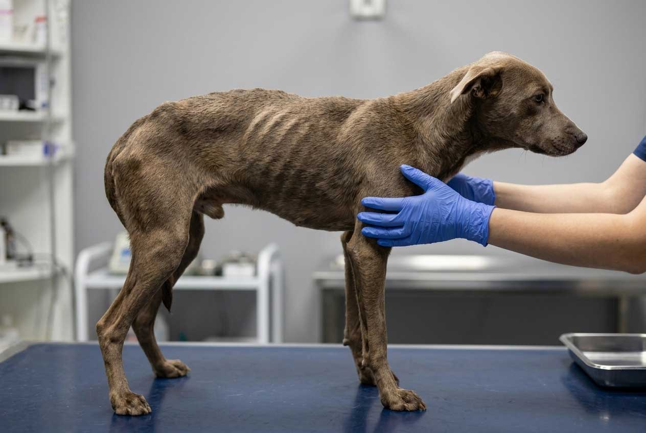
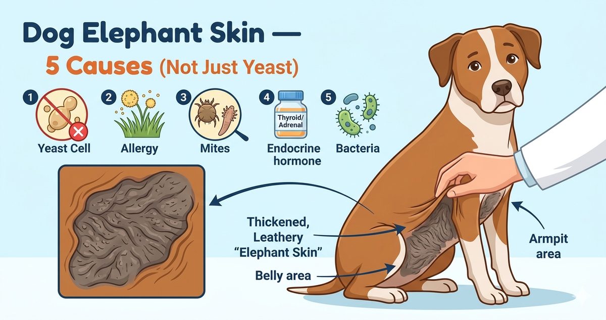
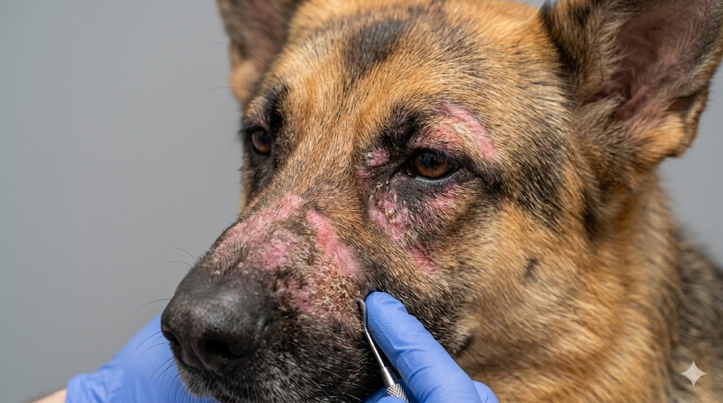
Elephant skin on dogs — medically known as lichenification — is the thick, darkened, leathery skin texture that develops after months of chronic inflammation, scratching, or irritation. Most quick guides will tell you the cause is yeast Malassezia dermatitis. That is the #1 cause but it is NOT the only one. Dog elephant skin has 5 distinct underlying causes (chronic yeast Malassezia, chronic atopic allergy, Cushing's disease and hypothyroidism, hyperkeratosis on nose or paw pads, and chronic mange), and the location plus other symptoms are the strongest clues to which cause is yours. This guide covers what causes elephant skin on dogs across all 5 causes, why lichenification in dogs sometimes does not go away even after treating the underlying trigger, how to distinguish Shar Pei wrinkled skin (genetic) from pathological elephant skin, and when to see a vet. For an instant AI photo check, our Dog Skin Black Spots Pictures AI tool identifies thickened darkened skin patterns from a photo.
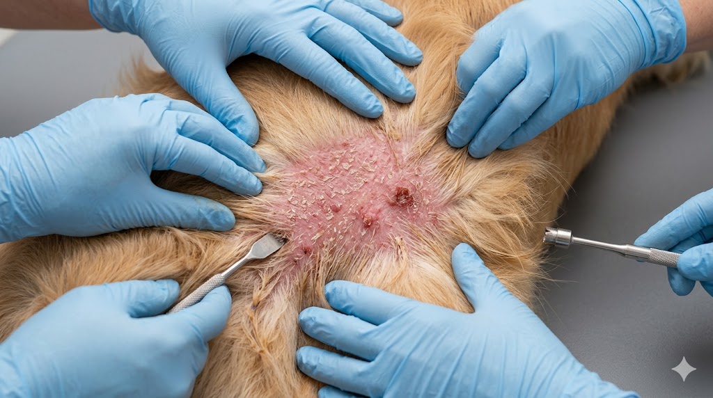
This article is for general educational purposes only and is not a substitute for professional veterinary diagnosis. Persistent dog elephant skin warrants a vet visit for proper differential workup including yeast cytology, allergy panels, and endocrine screening.
Want a faster differential? Our AI dog skin black spots pictures tool identifies thickened hyperpigmented patterns including elephant skin lichenification from a close-up photo.
Try Dog Skin Black Spots AI ToolWhy Does My Dog's Skin Look Like Elephant Skin?
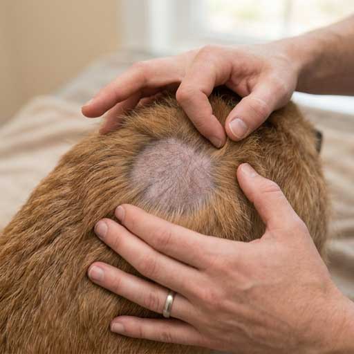
Why does my dog's skin look like elephant skin — the short answer is lichenification in dogs, the medical term for chronic skin thickening with darkened leathery texture. Lichenification develops in response to weeks or months of inflammation, scratching, rubbing, or skin barrier breakdown. The skin literally remodels itself in defense, becoming thicker and changing pigment. What causes elephant skin on dogs is rarely a single trigger — it is the cumulative end result of an underlying condition the dog has been dealing with for a long time. The 5 most common underlying causes are: (1) chronic yeast Malassezia dermatitis (the most common, but not the only), (2) chronic atopic allergy, (3) Cushing's disease or hypothyroidism, (4) hyperkeratosis on nose or paw pads, and (5) chronic mange. Identifying which of the 5 is yours requires looking at body distribution, smell, age, and other symptoms together.
Cause 1: Chronic Yeast Malassezia Dermatitis
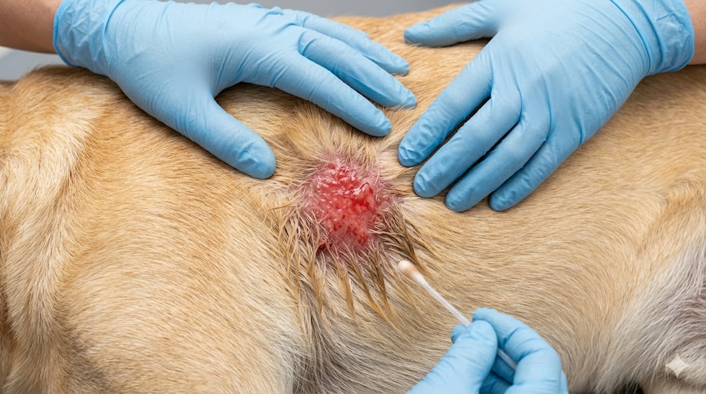
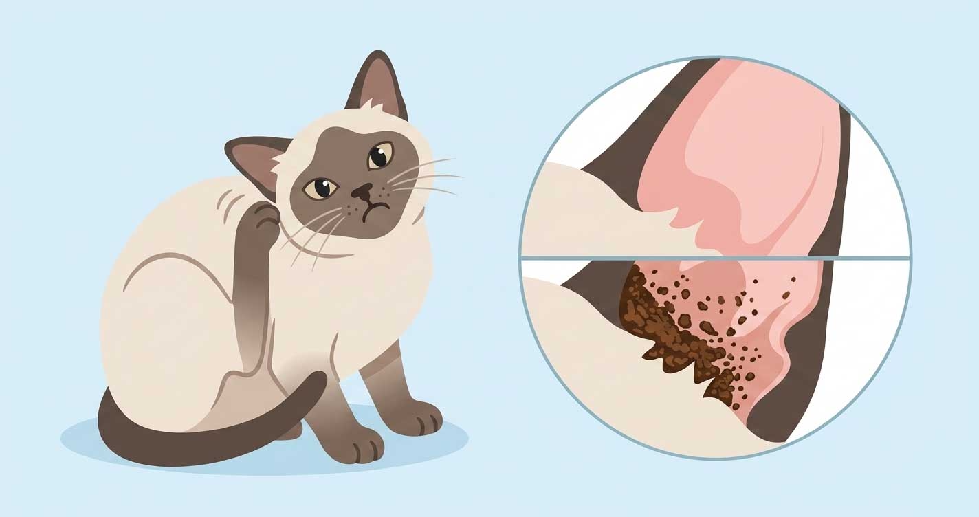
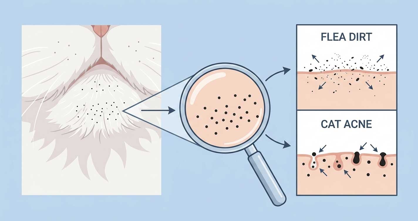
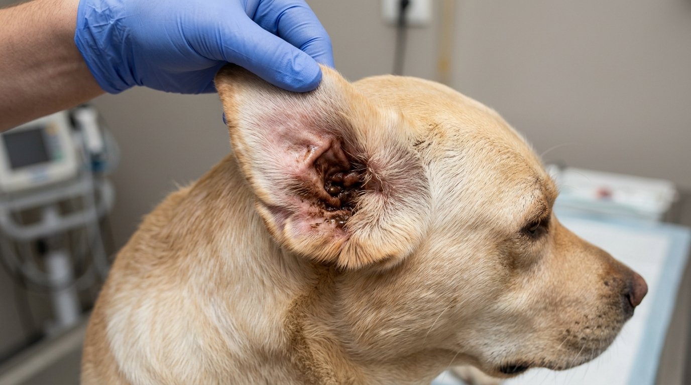
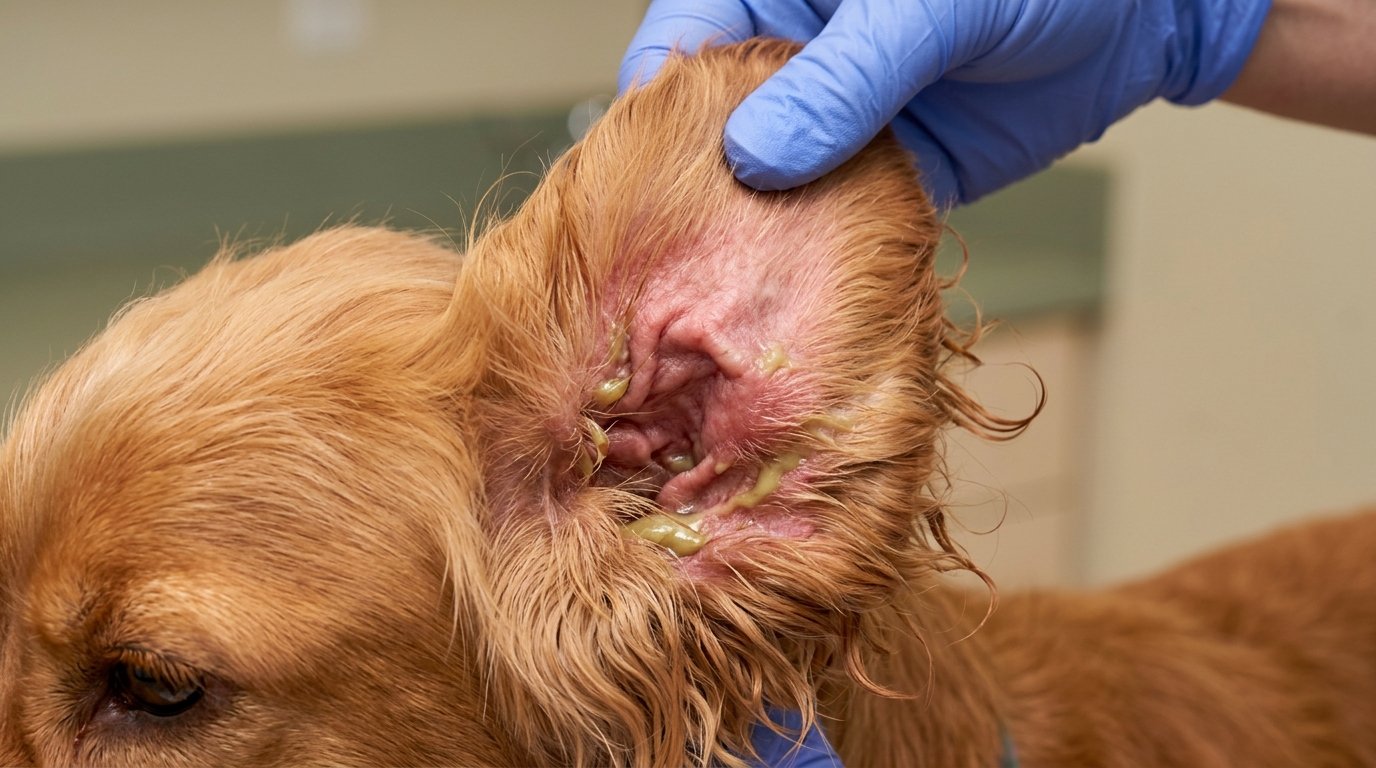
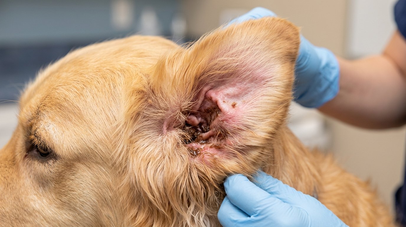
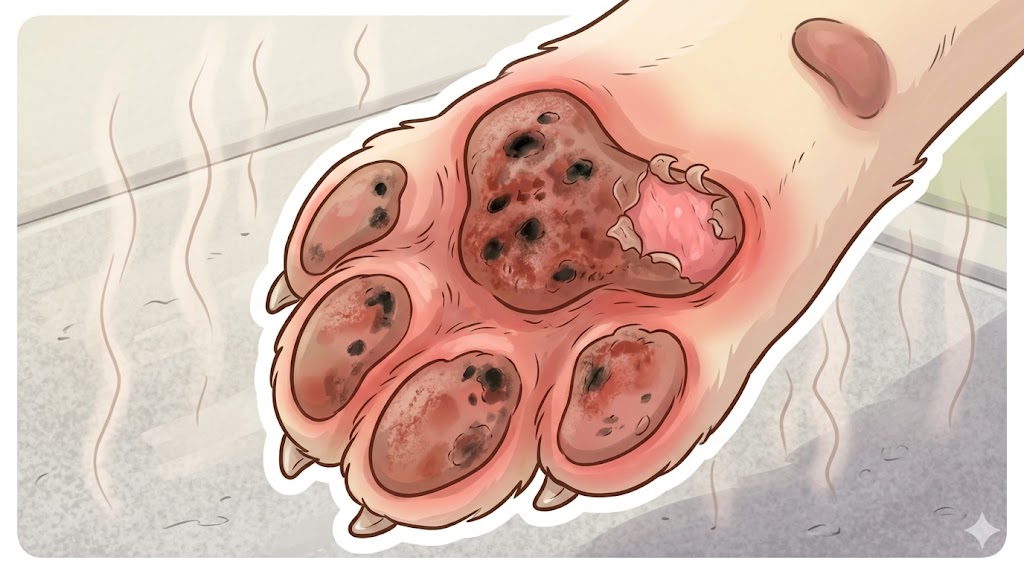
Chronic yeast Malassezia dermatitis is the most common cause of elephant skin on dogs and what most online sources point to first. Malassezia pachydermatis is a yeast that normally lives on dog skin in small numbers — when conditions allow it to overgrow (warm moist skin folds, chronic allergy, immune suppression), it triggers an extreme itch-scratch cycle that produces lichenification over weeks to months. Yeast dermatitis in dogs that progressed to elephant skin shows: (1) Black greasy thickened patches in skin folds (armpits, belly, groin, neck folds in brachycephalic breeds, between toes). (2) Sweet musty smell — often described as "corn chips" or "stale cheese." (3) Itching intensity varies from mild to extreme. (4) Often secondary to underlying atopic allergy — yeast is the proximate cause but allergy is the deeper root. The NIH PMC Canine Malassezia Dermatitis review confirms yeast is usually a secondary problem due to underlying skin disease, not a standalone primary cause.
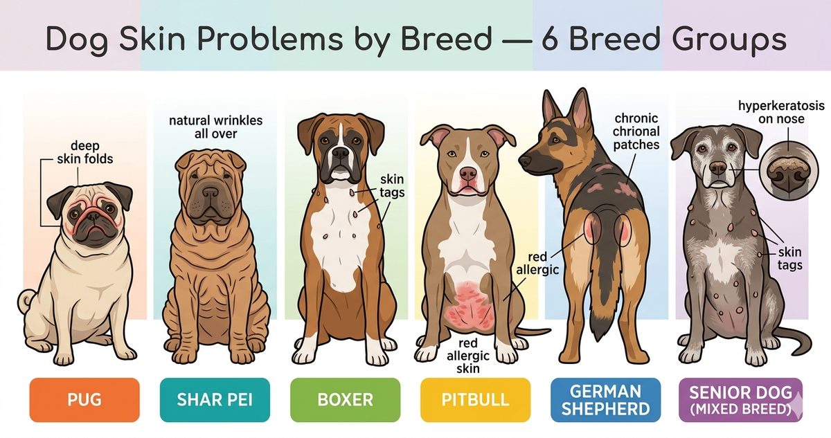
Body distribution signature: Skin folds + paws + ears, particularly in brachycephalic and folded-skin breeds (Bulldog, Pug, Shar Pei, Cocker Spaniel, Basset Hound). If your dog's elephant skin is concentrated in skin folds with a musty smell, malassezia dermatitis dog is the most likely underlying cause.
Cause 2: Chronic Atopic Allergy (Often the Deeper Root)
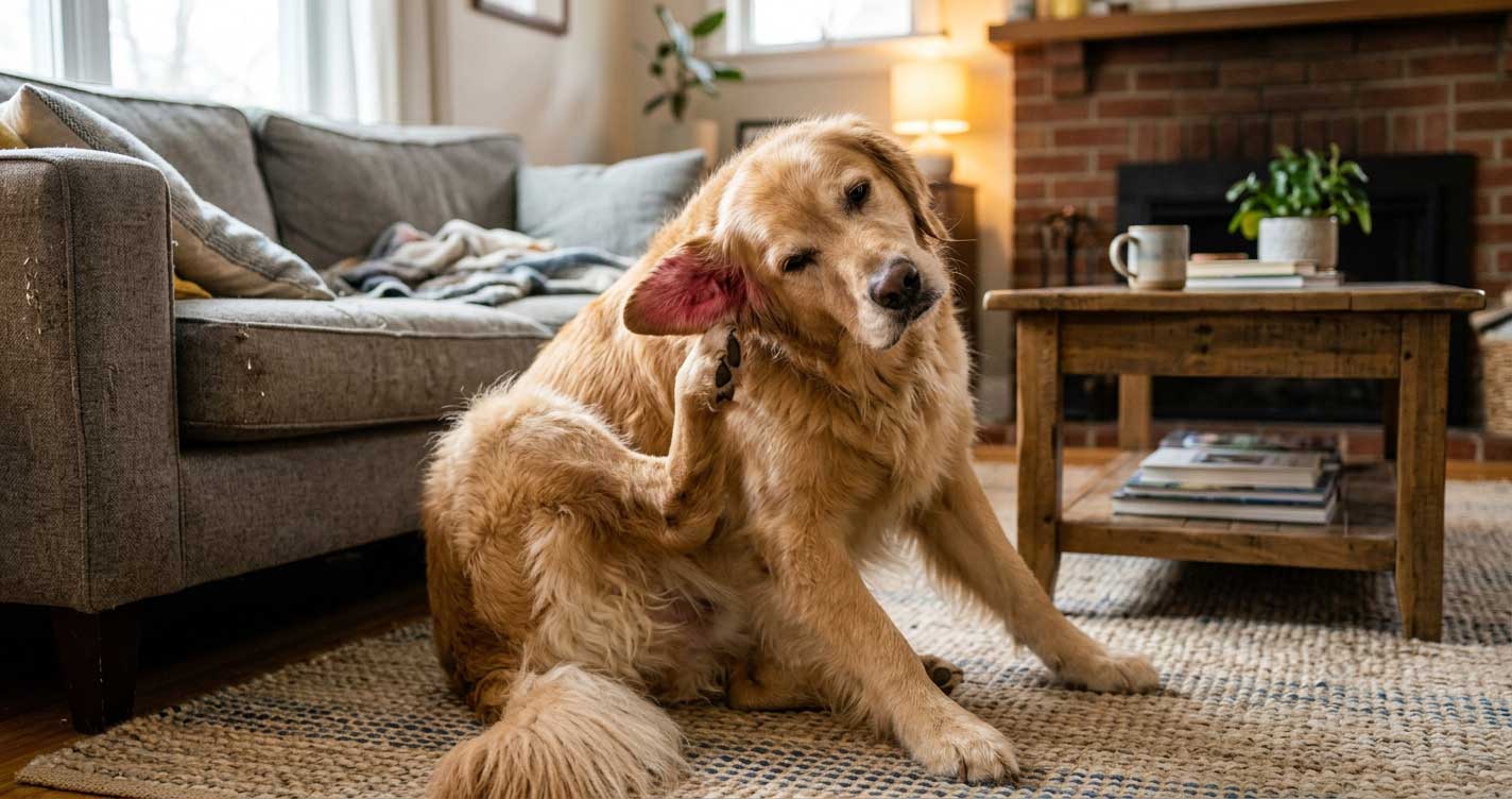
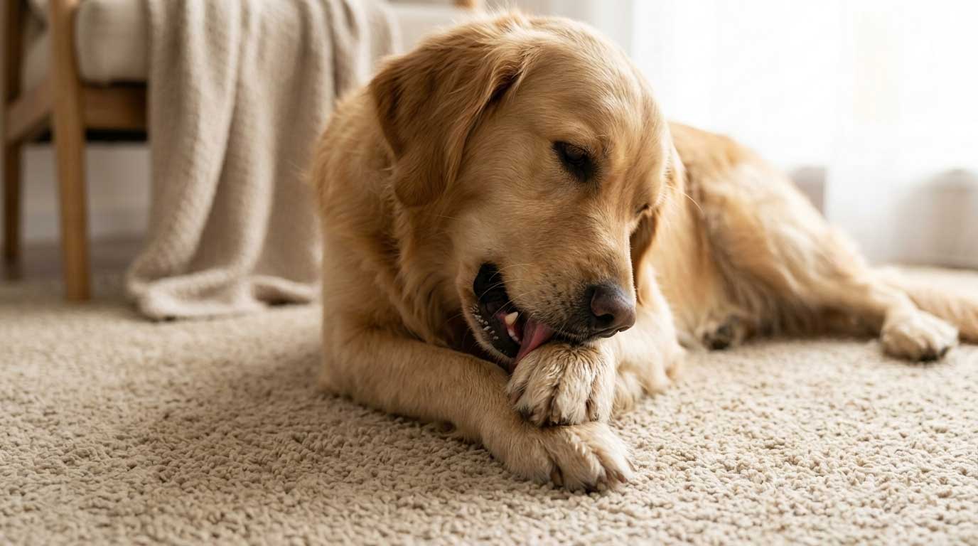
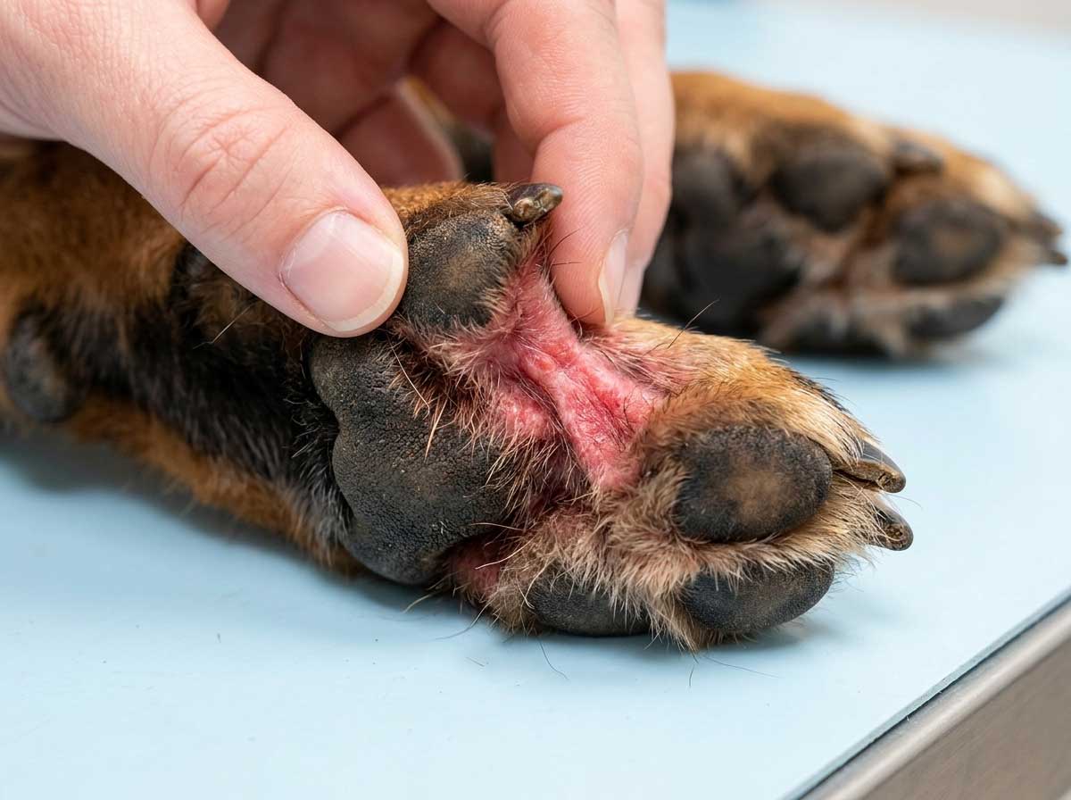
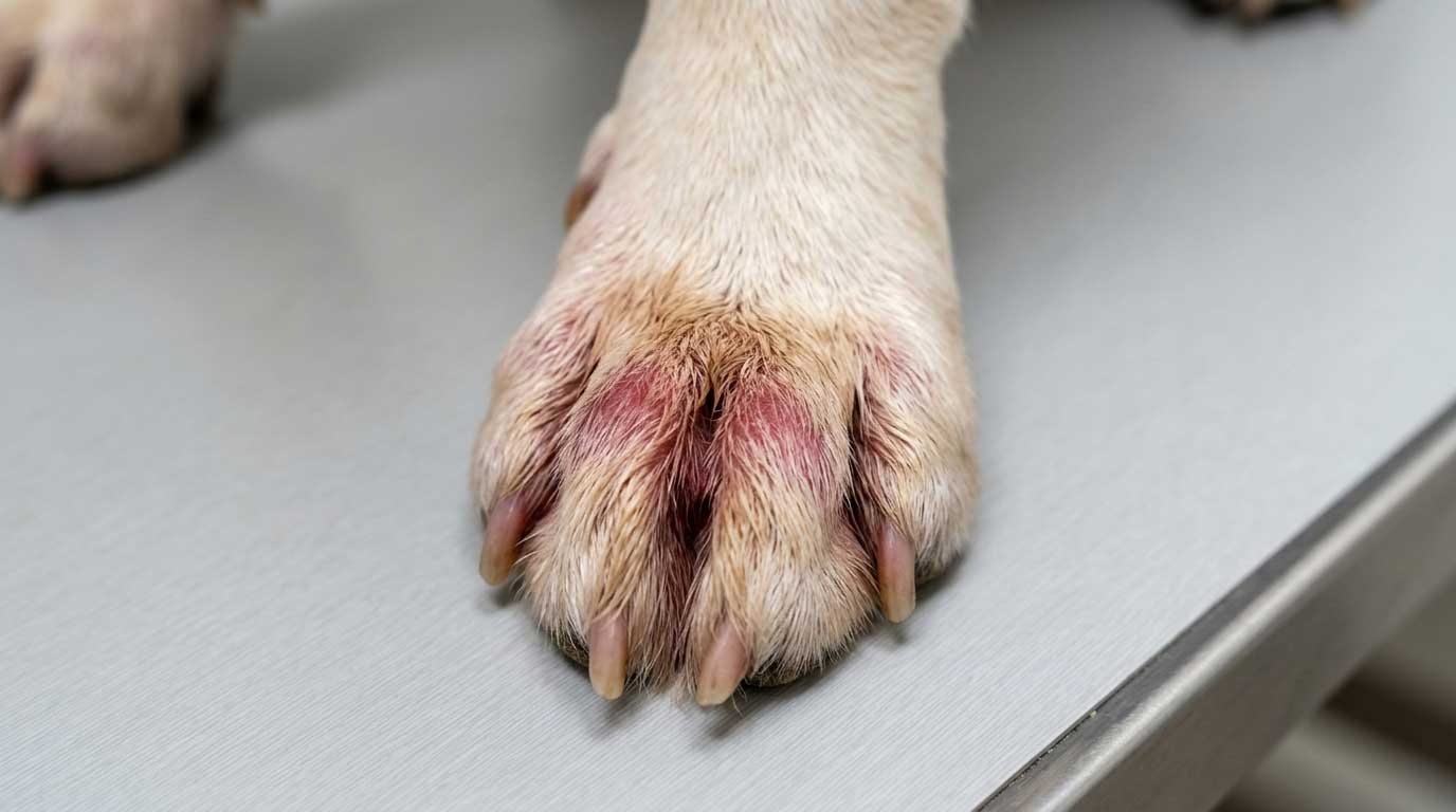
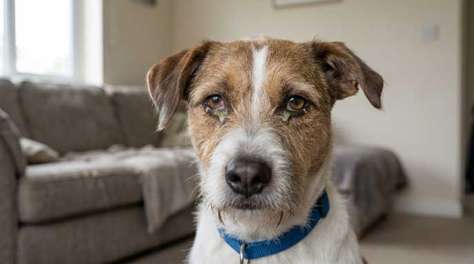
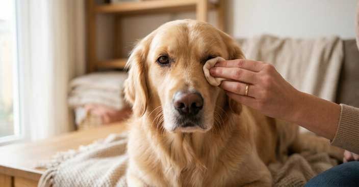
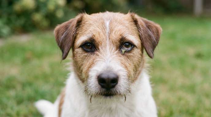
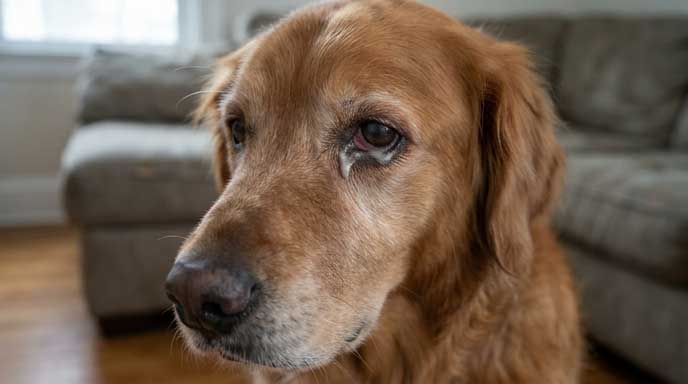
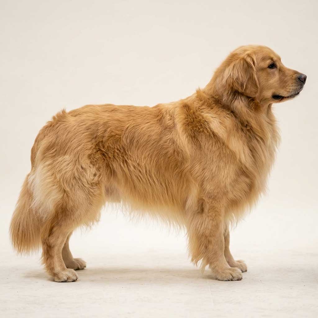
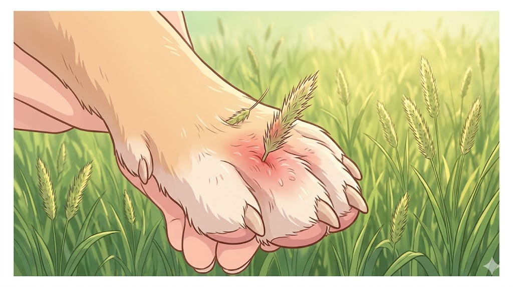
Chronic canine atopic dermatitis lichenification develops when atopic environmental allergy (pollen, dust mites, mold) has gone unmanaged for months. The dog scratches and chews target areas (paws, face, ears, armpits, groin), breaking the skin barrier and triggering remodeling. Even after the allergen is controlled, the thickened darkened skin often remains. Lichenification from atopic allergy shows: (1) Body distribution focused on paws (saliva staining on white paws), face, ears, armpits, and groin — NOT the lower back which is flea pattern. (2) Recurring ear infections without ear mites — the #1 sign of dog atopic dermatitis. (3) Seasonal worsening (spring and fall for pollen) or year-round (dust mites). (4) Often secondary yeast on top of atopic — the two interact and worsen the lichenification together. Certain breeds genetically predisposed include Golden Retrievers, Bulldogs, Labradors, German Shepherds, and West Highland Terriers.
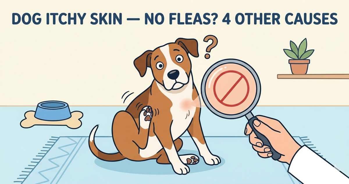
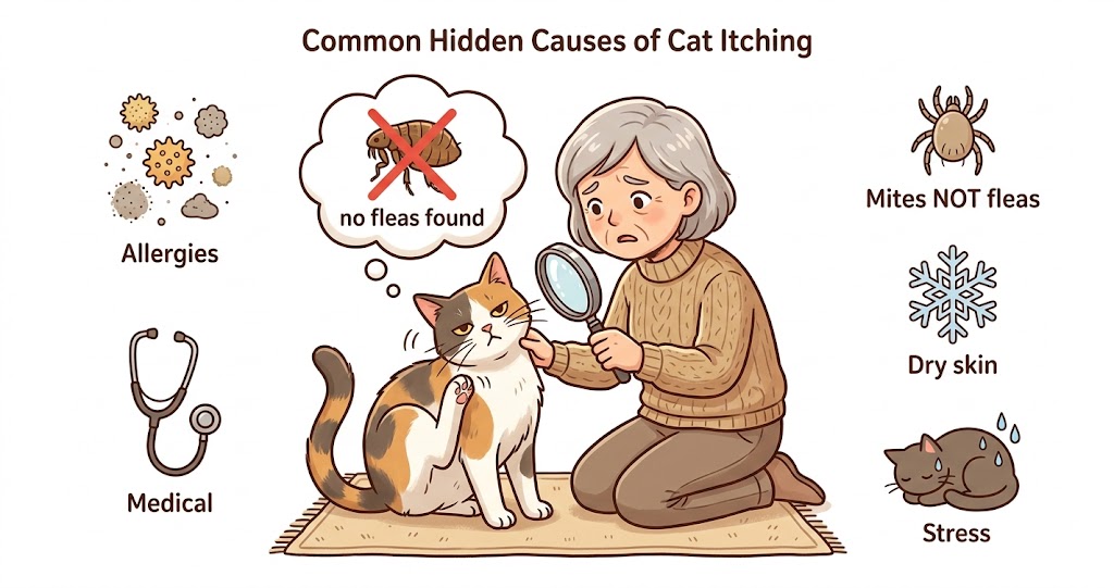
For deeper coverage of atopic allergy as the root cause of chronic itching that leads to elephant skin, our Dog Itchy Skin No Fleas 4 Causes guide covers atopic identification and differential vs other non-flea causes.
Chronic atopic allergy is the deeper root behind most elephant skin cases. Our dog itchy skin no fleas guide covers the 4 non-flea root causes including atopic dermatitis.
Read Dog Itchy Skin No Fleas GuideCause 3: Cushing's Disease and Hypothyroidism (Endocrine Causes)
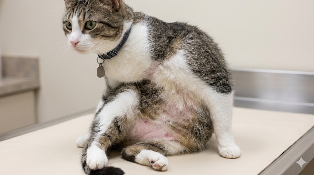
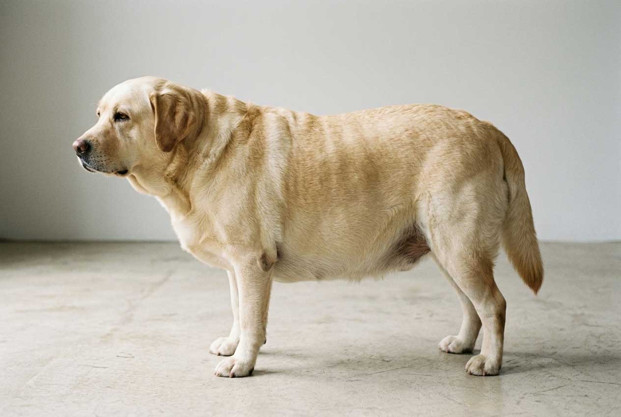
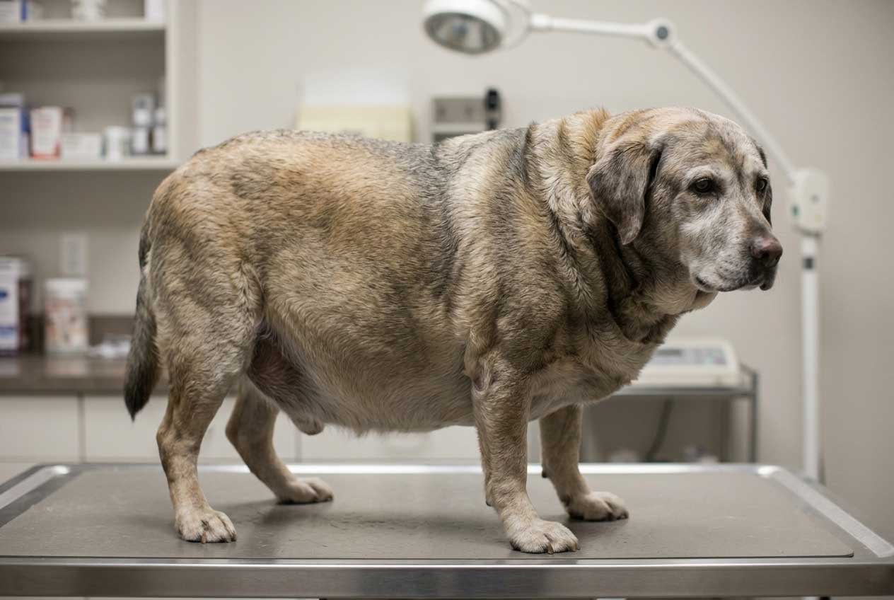
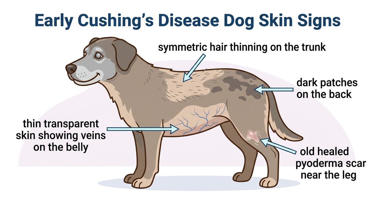
Cushing's disease skin thickening is a less commonly recognized but important cause of dog elephant skin. Cushing's (hyperadrenocorticism) is excessive cortisol production that causes systemic effects including widespread skin thinning followed by lichenification, hair loss, increased thirst and urination, pot-belly appearance, and panting. The skin in Cushing's often appears thin and bruised initially, but in chronic untreated cases progresses to thickened darkened patches. Hypothyroidism (under-active thyroid) causes a different but related skin pattern — dry coarse hair, hyperpigmentation, recurring skin and ear infections, and gradual lichenification on the back and trunk. Both endocrine causes share key differentiators from yeast or atopic: (1) Widespread rather than focal distribution. (2) Hair loss accompanying the skin change. (3) Behavioral or systemic signs (lethargy, increased thirst, weight changes). (4) Older dogs more affected (Cushing's peaks at 8-12 years, hypothyroidism most common 4-10 years).
Body distribution signature: Widespread lichenification + hair loss + systemic signs = endocrine workup needed. Among the 5 causes, Cushings disease skin thickening shares the most overlap with chronic yeast at first glance — both cause widespread thickened darkened skin — but Cushings adds pot-belly, thirst, and panting that yeast does not. The BluePearl Malassezia article and the VCA Yeast Dermatitis guide both flag endocrine disease as a differential when elephant skin is widespread rather than focal.
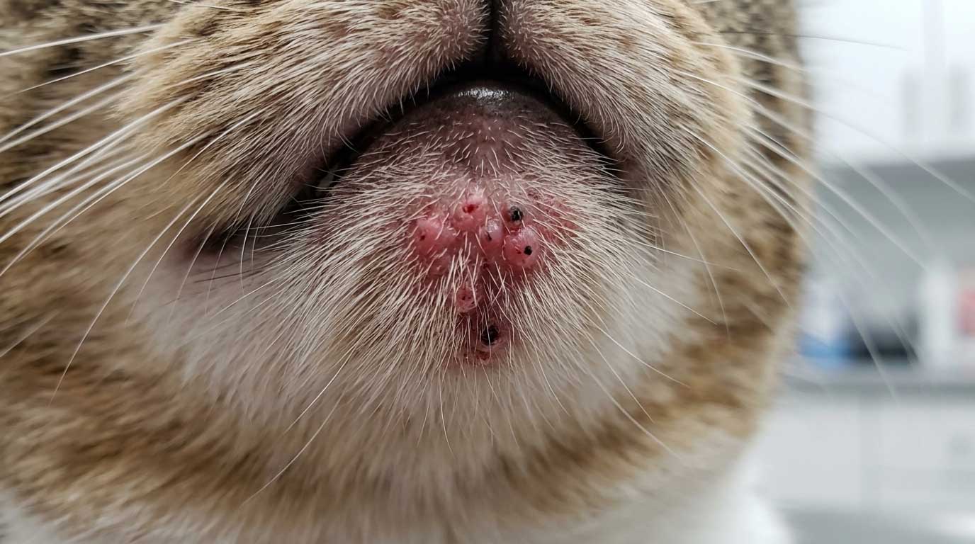
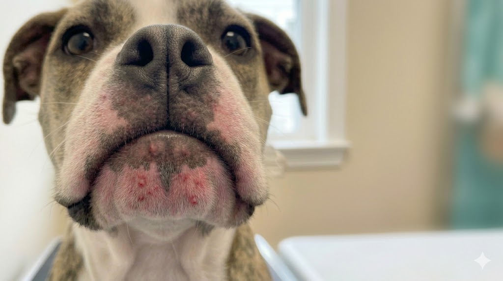
Cause 4: Hyperkeratosis (Nose or Paw Pad — Localized)
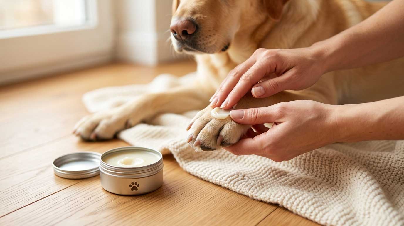
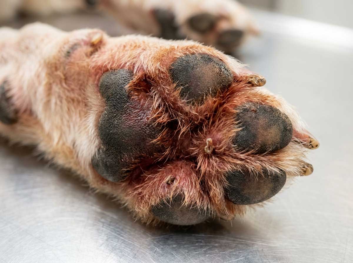
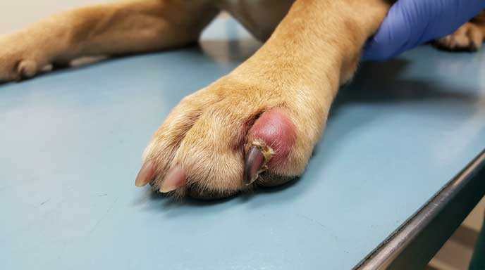
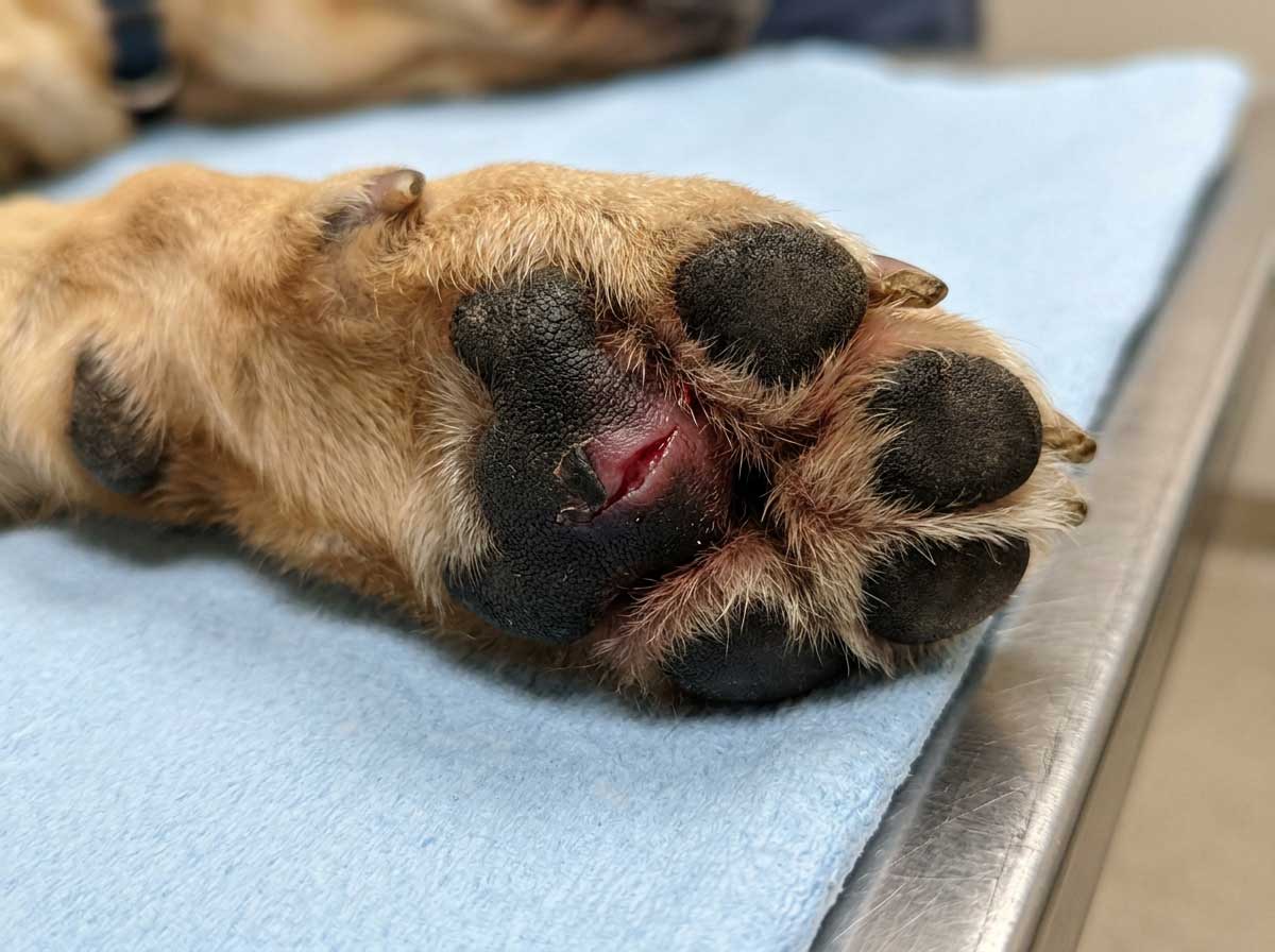
Hyperkeratosis nose paw pad dog is a distinct cause of elephant skin pattern that affects only the nose leather (nasal hyperkeratosis) or paw pad surfaces (digital hyperkeratosis). Unlike systemic lichenification from yeast or allergy, hyperkeratosis is a localized condition where the outer skin layer overgrows and hardens. Visual signs: (1) Crusty, hard, sometimes cracking surface on the nose or paw pads only. (2) Other body areas completely normal. (3) Common in senior dogs (8+ years) as an aging-related change. (4) Some breeds have a genetic predisposition (Dogue de Bordeaux, Bedlington Terrier, Labrador). (5) Idiopathic hyperkeratosis (no underlying disease) is common in seniors and considered cosmetic rather than dangerous. (6) Secondary hyperkeratosis can develop from underlying conditions (autoimmune disease, distemper history, zinc deficiency in certain breeds). The key differentiator from other elephant skin causes: location is restricted to nose / paw pads only, dog otherwise comfortable, often slow gradual onset over months.
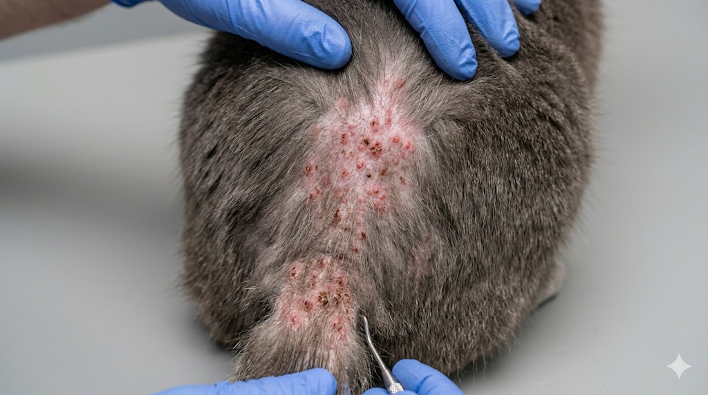
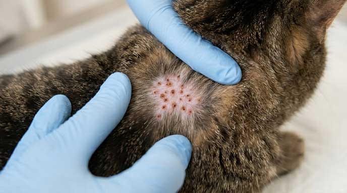
Cause 5: Chronic Mange (Demodex Generalized)
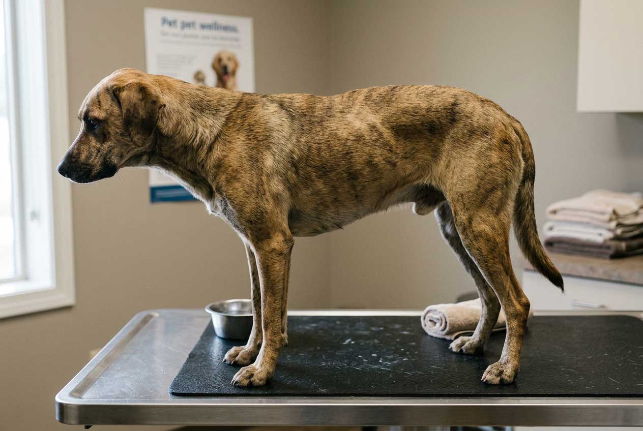
Chronic mange thickened skin dog patterns develop when long-untreated demodectic mange (generalized Demodex canis) causes severe inflammation that remodels the skin over months. Sarcoptic mange acute cases usually present as extreme itching with crusty red skin before lichenification has time to develop — but generalized demodicosis in immunocompromised dogs (especially puppies and seniors with endocrine disease) can produce widespread elephant skin over time. Mange-driven lichenification shows: (1) Widespread distribution + bald patches. (2) History of skin scrapings positive for Demodex. (3) Often associated with underlying immune compromise (Cushing's, hypothyroidism, immunosuppressive therapy). (4) Young puppies (juvenile-onset Demodex) can develop generalized lichenification if untreated. The differential from atopic + yeast: mange typically shows on skin scraping cytology, atopic + yeast do not.
Shar Pei Wrinkled Skin vs Elephant Skin — How to Tell Apart
Shar pei wrinkled skin vs elephant skin is a critical visual differential because Shar Pei dogs have genetic wrinkled skin from birth, which can look concerning but is NOT pathological lichenification. Genetic Shar Pei wrinkles show: (1) Present from puppyhood. (2) Even, symmetric distribution. (3) Skin color matches surrounding normal coat. (4) No active inflammation, scratching, or smell. (5) Hair coat normal. (6) Common in juvenile dogs of the breed. Pathological elephant skin in a Shar Pei (or any breed) shows: (1) New darker patches developing after months. (2) Skin smells musty or yeasty. (3) Active scratching or licking. (4) Hair loss on the thickened areas. (5) Asymmetric distribution. (6) Other body areas may have rash, redness, or sores. Owners of Shar Pei especially need to watch for chronic yeast hidden in the wrinkles — the breed's anatomy creates ideal yeast habitat. The same applies to other folded-skin breeds (Bulldog, Pug, Bullmastiff, Bloodhound).
Does Elephant Skin on Dogs Go Away?
Does elephant skin on dogs go away — this is one of the most common owner questions, and the honest answer is mixed. Early stage lichenification (caught within weeks of starting) often reverses fully once the underlying cause is controlled. Late stage lichenification (months to years of unmanaged inflammation) often remains as permanent darkened thickened skin even after the trigger is resolved. The factors that determine reversibility: (1) How long the inflammation has been present (under 2 months = often reversible, over 6 months = often permanent). (2) How severe the chronic scratching has been. (3) Whether the underlying cause can be fully controlled. (4) The dog's age and overall skin healing capacity. For owners of dogs with established elephant skin: even if the texture does not fully reverse, controlling the underlying cause prevents further progression and stops new patches from forming. The visible lichenification becomes "cosmetic" rather than active disease. The VCA Yeast Dermatitis in Dogs guide covers expected outcomes for yeast-driven cases.
Pictures of Elephant Skin on Dogs — Visual Reference Patterns
Pictures of elephant skin on dogs show distinct visual patterns by underlying cause. Whether you searched "elephant skin dog" (singular case) or "elephant skin dogs" (across cases), the 5-cause matrix below summarizes the differential visual signs that apply across all individual cases:
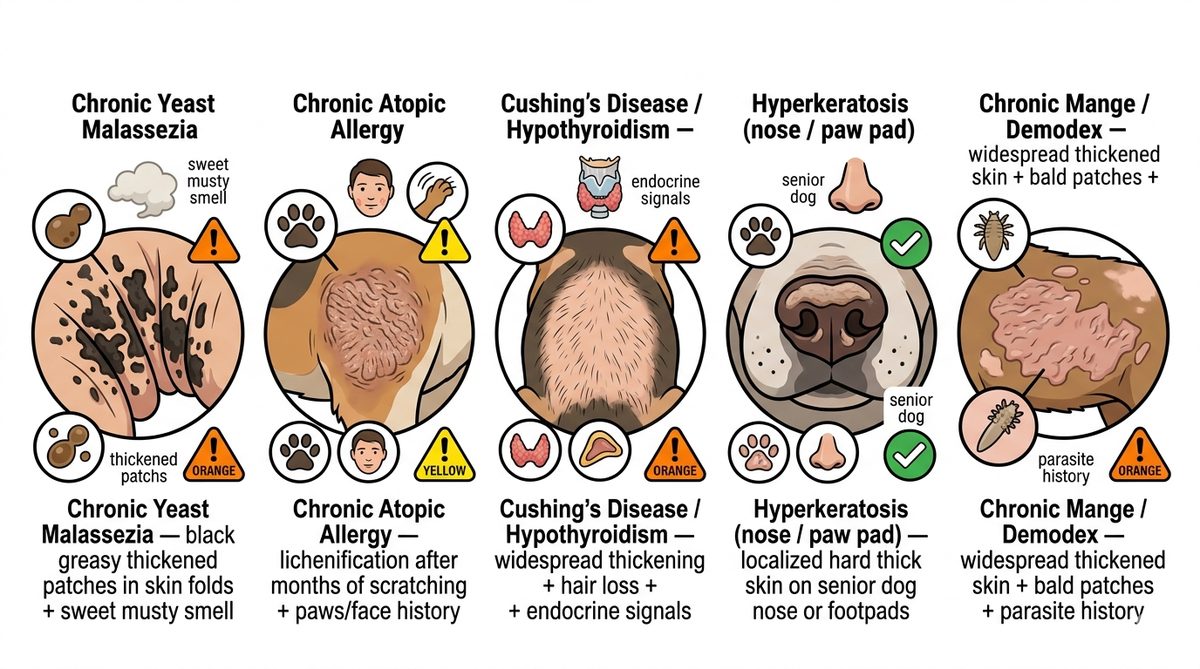
- ✓**Chronic Yeast Malassezia** — Black greasy patches in folds + sweet musty smell + recurring itch (most common)
- ✓**Chronic Atopic Allergy** — Paws/face/ears focus + saliva staining + recurring ear infections + seasonal pattern
- ✓**Cushing's / Hypothyroidism** — Widespread + hair loss + increased thirst + pot belly + senior dog
- ✓**Hyperkeratosis** — Nose or paw pads only + crusty hardened surface + dog otherwise normal
- ✓**Chronic Mange (Demodex)** — Widespread + bald patches + history of mites + immune compromise
When to See a Vet — Decision Framework
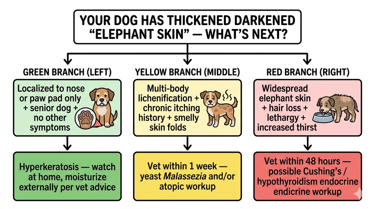
Use this dog elephant skin vet decision framework based on distribution + accompanying symptoms:
- ✓**Watch at home (or routine vet)**: Hyperkeratosis on nose or paw pads only + senior dog + no itching or smell + no other symptoms
- ✓**Vet within 1 week**: Multi-body lichenification + chronic itching history + skin folds smell musty or yeasty + recurring ear infections
- ✓**Vet within 48 hours**: Widespread elephant skin + hair loss + increased thirst and urination + pot-belly appearance + lethargy — possible Cushing's or hypothyroidism endocrine workup needed
- ✓**Always vet (do not just watch)**: New rapidly developing thickening + skin breakdown or open sores + worsening despite at-home care + young dog with widespread pattern (juvenile demodicosis suspected)
Before your vet visit, prepare these data points: (1) When did you first notice the thickening? (2) Has the dog been scratching specific areas? (3) Body distribution (focal vs widespread). (4) Any smell from affected skin? (5) Recent appetite, thirst, energy changes. (6) Recent ear infections or paw chewing. (7) Other dogs / pets affected.
Dog elephant skin focused on belly or belly fold? Our AI dog belly rash pictures tool identifies yeast vs allergy vs FAD vs bacterial pattern in belly distribution.
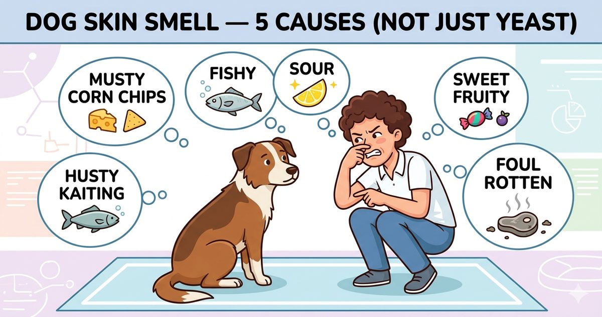
Try Dog Belly Rash AI ToolChronic yeast that turned skin black also smells musty. Our new dog skin smell guide covers 5 distinct smells (yeast / fishy / sour / sweet fruity emergency / foul) with differential.
Read Dog Skin Smell 5 Causes GuideRelated Reading on Dog Skin Conditions
Deeper guides on related dog elephant skin lichenification topics: Dog Skin Allergy — 3 Types and What Helps covers atopic + food allergy as the chronic root behind many lichenification cases; Dog Itchy Skin No Fleas 4 Causes covers the chronic scratching that leads to elephant skin; Dog Paw Yeast Infection covers paw-specific Malassezia patterns. For an instant AI photo differential, our Dog Skin Black Spots Pictures AI tool identifies thickened darkened skin patterns from a photo of your dog.
Frequently Asked Questions
Why does my dog's skin look like elephant skin?
+
Does elephant skin on dogs go away?
+
What causes elephant skin on dogs besides yeast?
+
How can I tell Shar Pei wrinkled skin vs pathological elephant skin?
+
Is elephant skin on dogs the same as hyperpigmentation?
+
Pictures of elephant skin on dogs — what do they look like?
+
Dog Elephant Skin — Need a Faster Differential?
Upload a clear photo of your dog's thickened darkened skin for an instant AI differential between yeast vs atopic vs endocrine vs hyperkeratosis vs mange — beyond what AI Overview tells you.
Disclaimer: This article is for informational purposes only and is not a substitute for professional veterinary advice. Always consult a licensed veterinarian for diagnosis and treatment of your pet's health conditions.